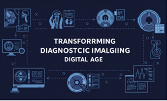
Introduction to DIAG Image
DIAG Image When it comes to modern medicine, DIAG Image, or diagnostic imaging, is nothing short of revolutionary. It allows doctors to see inside the human body without making a single incision. Think of it as the eyes of modern healthcare—detecting disease, monitoring treatment, and guiding surgery, all without opening up the patient.
But what exactly is a DIAG Image? Simply put, it’s a visual representation of the interior of the body created by various technologies like X-rays, CT scans, MRIs, and more. These images are used to diagnose conditions, guide medical procedures, and evaluate the effectiveness of treatments.
Evolution of Diagnostic Imaging
From X-rays to AI-integrated scans
The journey began with the discovery of X-rays in the late 19th century. Fast forward to today, and we have AI-enhanced 3D imaging that can detect diseases even before symptoms appear.
Major Milestones in Diagnostic Imaging
-
1895 – Wilhelm Roentgen discovers X-rays
-
1971 – First CT scan performed
-
1980s – MRI technology becomes commercialized
-
2020s – AI integration becomes a standard in imaging analysis
Types of DIAG Imaging Technologies
X-Ray Imaging
This is the oldest and still most widely used DIAG image technique. It’s great for spotting fractures, infections, and tumors.
CT Scans
CT, or computed tomography, provides detailed cross-sectional images. It’s like X-ray’s smarter sibling—able to create 3D visuals.
MRI Scans
MRIs use magnetic fields and radio waves, offering unparalleled detail of soft tissues. Neurologists and orthopedics love it.
Ultrasound Imaging
Safe for pregnant women and quick diagnoses, ultrasound uses sound waves to produce images in real-time.
PET Scans
Want to know how your organs are functioning? PET scans visualize metabolic activity and are often used in cancer diagnosis.
Applications of DIAG Image in Healthcare
Diagnostic imaging isn’t just about “seeing” inside the body—it’s about understanding what’s going wrong and where. Whether it’s spotting a tiny tumor or ensuring a stent is placed correctly, DIAG images are indispensable.
-
Early Detection – Spotting conditions before symptoms appear
-
Treatment Monitoring – Tracking progress in diseases like cancer
-
Surgical Planning – Helping doctors navigate complex anatomy
The Role of AI in DIAG Image
Welcome to the future—AI doesn’t just assist doctors; it learns from millions of images to enhance diagnostic accuracy. Machine learning algorithms can flag abnormalities, assist with prioritization, and reduce human error.
Imagine an algorithm that can read a chest X-ray faster than a trained radiologist—it’s happening.
Benefits of Diagnostic Imaging
Why has DIAG imaging become a pillar of healthcare?
-
Non-invasive: No cutting, no bleeding.
-
Speed: Immediate results in emergency cases.
-
Precision: Helps in targeting treatments exactly where needed.
-
Versatility: Used in everything from dentistry to neurosurgery.
DIAG Image in Different Medical Fields
Oncology
Detect tumors early and monitor the effect of chemotherapy.
Neurology
Scan the brain for signs of stroke, tumors, or degenerative diseases.
Cardiology
Visualize arteries, valves, and heart function.
Orthopedics
Diagnose fractures, ligament tears, and joint conditions.
Challenges and Limitations
Nothing is perfect, not even DIAG imaging.
-
Radiation Risks – Especially with CT scans.
-
High Costs – Equipment and maintenance aren’t cheap.
-
False Positives – Can lead to unnecessary procedures.
Recent Innovations in DIAG Imaging
-
3D Imaging – Perfect for surgical planning and patient education.
-
Portable Devices – Handheld ultrasound machines now exist.
-
Cloud PACS – Doctors can view your scans anywhere in the world.
Future Trends in DIAG Imaging
The future looks like something out of a sci-fi movie:
-
AI at the bedside: Immediate analysis as scans are performed.
-
Real-Time Surgery Imaging: Visual updates during surgery.
-
Customized Diagnostics: Imaging that adapts to your genetics.
Ethical and Legal Considerations
With great data comes great responsibility.
-
Privacy – Who owns your scans?
-
AI Bias – Algorithms trained on limited datasets could miss diagnoses in minority groups.
DIAG Image in Telemedicine
COVID-19 made remote healthcare a necessity, and DIAG image tech stepped up.
-
Cloud Sharing – Instantly share images with specialists.
-
Mobile Access – View scans on tablets or smartphones.
-
Remote Diagnosis – No need for the doctor and patient to be in the same room.
Conclusion
In a world where seeing is believing, DIAG images are the eyes through which medicine views the human body. From life-saving diagnoses to real-time surgical guidance, diagnostic imaging has revolutionized how healthcare works. As technology advances—especially with AI and cloud computing—this field is only going to get sharper, smarter, and more accessible.
❓FAQs
1. What is the most common DIAG image type used today?
X-rays remain the most frequently used due to their affordability and speed.
2. Are DIAG images safe?
Most are safe, though some like CT scans involve radiation. Always consult with your physician.
3. How is AI changing diagnostic imaging?
AI helps interpret images faster and with higher accuracy, often assisting radiologists in identifying issues sooner.
4. Can DIAG images detect all types of cancer?
No, but combined imaging techniques (like PET + CT) significantly improve detection accuracy.
5. How do I prepare for a diagnostic imaging procedure?
It depends on the scan type. Some require fasting or contrast agents. Always follow the instructions provided by your doctor.

